
Dental X-rays are information-dense images, but they are not naturally patient-friendly. A dentist can identify an incipient caries lesion or quantify periodontal bone loss while a patient sees little more than a grayscale image.
That communication gap is where AI dental imaging companies 2024 became particularly interesting. Platforms from companies such as Pearl, Overjet, VideaHealth and Diagnocat increasingly used computer vision to analyze radiographs, highlight findings and create visual explanations that clinicians could use during consultations. A 2024 review of AI in dentistry identified Overjet, Pearl and VideaHealth among the commercial systems receiving regulatory clearances for dental AI applications.
The important distinction is that these systems are not simply “AI dentists.” They are generally clinical decision-support and visualization systems. The clinician remains responsible for interpreting findings, confirming the diagnosis and deciding whether treatment is appropriate.
The most useful way to evaluate the 2024 landscape, therefore, is to follow the data pipeline: capture the image, enhance it, analyze it, annotate clinically relevant regions, review the output and then translate those findings into patient-friendly education.
What Are AI Dental Imaging Companies?
AI dental imaging companies develop software that applies machine learning and computer vision to dental radiographs, photographs, CBCT scans or other clinical images.
A typical system does not simply classify an X-ray as “healthy” or “unhealthy.” Depending on its intended use, it may detect suspected caries, identify bone-level changes, locate anatomical structures, segment teeth or enhance image quality.
This matters for patient education because raw radiographs can be difficult for non-clinicians to interpret. AI-generated overlays can transform subtle visual patterns into more understandable evidence.
A 2024 British Dental Journal report described AI tools integrated into Planmeca Romexis as supporting dental imaging, diagnosis, treatment planning and patient communication.
How Does AI Dental Imaging Work?
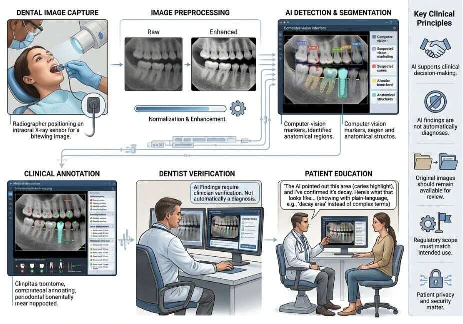
Most dental imaging systems can be understood as a multi-stage computer-vision pipeline.
1. Image acquisition
The workflow begins with an intraoral X-ray, bitewing, panoramic image, cephalometric image or 3D scan.
The quality and modality of the input matter because a model trained for one image type should not automatically be assumed to perform the same task on another.
2. Image preprocessing
Preprocessing can improve contrast, normalize image characteristics or reduce visual noise.
Overjet’s 2024 introduction of IRIS, for example, described an AI-native imaging workflow combining image capture, enhancement, analysis and explanation.
3. Detection and segmentation
The model searches for learned patterns associated with specific clinical findings.
Segmentation goes further by identifying the boundaries of structures or lesions rather than simply assigning a label.
4. Annotation
The output can become a colored overlay, measurement, tooth map or other visualization.
This is arguably the most important layer for patient education because the system converts an abstract prediction into something the patient can see.
5. Human review
The dentist evaluates the AI output against the original image, clinical examination and patient history.
6. Patient presentation
Finally, the clinician uses the visual evidence to explain:
- what was detected;
- where it is located;
- how extensive it appears;
- why it matters;
- what treatment or monitoring options exist.
Architect’s Note: The best patient-education workflow does not hide the original radiograph. AI annotations should be presented as an additional evidence layer so the clinician can compare the model output with the source image.
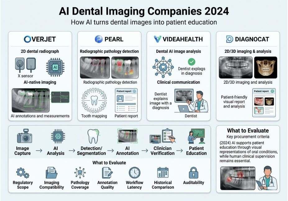
AI Dental Imaging Companies 2024: 4 Notable Platforms
The 2024 market included several companies with different strengths. They should not be treated as interchangeable because regulatory status, supported modalities, pathology detection and patient-facing capabilities vary.
| Company / platform | Primary imaging focus | Patient-education capability | Important evaluation point |
| Overjet | 2D dental radiographs and AI-native imaging | Color annotations, measurements, image comparison | Strong emphasis on integrated imaging and clinical visualization |
| Pearl | Dental radiograph analysis | Color-coded findings, tooth mapping, reports | Strong chairside communication and pathology visualization |
| VideaHealth | Dental AI image analysis | Visual findings supporting clinical conversations | Evaluate exact cleared use cases and integrations |
| Diagnocat | 2D and 3D dental imaging | Patient-friendly reports and visual tooth charts | Particularly relevant where 3D analysis and reporting matter |
A peer-reviewed review of dental AI applications lists Diagnocat, Denti.AI, Videa.AI, Overjet, CephX, DentalXR.ai and Pearl among evaluated commercial applications, with services ranging from 2D/3D imaging to decay and bone-loss detection.
Overjet
Overjet is notable for combining dental AI with an imaging environment. Its 2024 IRIS announcement positioned AI directly inside the imaging workflow rather than as a separate application.
Its approach emphasizes image enhancement, AI findings, measurements and patient presentation.
Pearl
Pearl’s Second Opinion platform focuses heavily on radiographic pathology detection and chairside communication. Its current product materials describe color-coded findings, tooth-part mapping, comparison views and patient health reports.
Its patient-education value comes from making the AI output visually interpretable rather than requiring the patient to understand radiographic anatomy.
VideaHealth
VideaHealth was another significant 2024 player. Becker’s reported that VideaHealth received FDA clearance for Videa Dental Assist in 2024.
For practices evaluating VideaHealth or similar platforms, the key question is not simply whether a product is “FDA cleared,” but which specific functions and patient populations the clearance covers.
Diagnocat
Diagnocat takes a broader imaging approach, including 2D and 3D analysis. Its patient-report workflow uses simplified language and visual representations, positioning AI analysis as a communication layer as well as a clinical-support tool.
Pro Tip: Build your shortlist by clinical task not brand recognition. A practice that primarily uses bitewing radiographs has different requirements from an oral-surgery or implant practice relying heavily on CBCT.
How AI Improves Dental Patient Education
The strongest patient-education use case is visual grounding.
Instead of telling a patient, “You have early interproximal decay,” a dentist can point to the relevant region on an annotated radiograph.
The 2024 Cureus review found that visual representation of dental problems can encourage patients to understand their oral condition, adopt healthier practices and seek timely treatment.
Useful patient-education features include:
- Color-coded findings that identify suspicious regions.
- Measurements that communicate severity more concretely.
- Tooth mapping that connects a finding to a specific tooth.
- Side-by-side comparisons that show progression.
- Plain-language reports that reduce technical terminology.
- Historical image comparison that helps patients understand change over time.
The communication model becomes:
Clinical finding → visual evidence → plain-language explanation → patient question → informed decision
That is considerably more useful than simply showing an unprocessed radiograph.
Why this matters
Patients often do not have the training to distinguish an important radiographic finding from normal anatomical variation.
AI does not solve that communication problem by itself. The value comes from combining algorithmic visualization with clinician explanation.
What Should Practices Compare Before Choosing a Tool?
A feature checklist is not enough. Practices should evaluate the entire clinical and technical workflow.
1. Regulatory scope
Check the actual intended use and regulatory clearance rather than relying on a vendor’s general “FDA-cleared AI” statement.
2. Supported imaging
Determine whether the platform supports your existing:
- intraoral sensors;
- panoramic systems;
- CBCT;
- imaging software;
- practice-management system.
3. Pathology coverage
Ask which findings the model is designed and validated to detect.
4. Annotation quality
Patient education depends heavily on whether annotations are understandable without becoming visually distracting.
5. Workflow latency
A model that takes too long to generate results can disrupt chairside communication.
6. Historical comparison
Longitudinal visualization can be more educational than a single snapshot because it shows whether a finding has changed.
7. Auditability
Clinicians should be able to distinguish the original image from AI-generated overlays and understand what the system actually reported.
8. Privacy and security
Dental radiographs are patient health information. Evaluate encryption, access controls, retention policies and contractual data handling.
Technical Note: Treat regulatory clearance, clinical validation and cybersecurity as three separate evaluation dimensions. A product can perform well technically without being cleared for every clinical use a salesperson might discuss.
Step-by-Step: Build an AI Patient-Education Workflow
A practical deployment does not require an autonomous agent. A deterministic pipeline is often safer.
Step 1: Capture
Acquire the patient’s radiograph using the practice’s normal imaging workflow.
Step 2: Analyze
Send the image through the selected AI system according to its supported workflow.
Step 3: Verify
The dentist compares AI findings against the original image and clinical examination.
Step 4: Annotate
Select only findings that are clinically relevant to the conversation.
Step 5: Explain
Use the image to explain the condition in plain language.
Step 6: Document
Record the clinical assessment and treatment decision separately from the AI output.
A simplified software abstraction might look like this:
def patient_education_pipeline(radiograph, clinical_context):
    image = preprocess(radiograph)

    ai_findings = dental_ai.analyze
        image=image,
        allowed_use_cases=["caries", "bone_loss"]


    verified_findings = clinician_review
        image=image,
        findings=ai_findings,
        context=clinical_context


    patient_view = create_visual_summary
        image=image,
        findings=verified_findings,
        language="plain"


    return patient_view
The important design principle is the clinician_review() stage. An implementation should not automatically convert a model prediction into a diagnosis or treatment recommendation.
Common Mistakes and How to Avoid Them
Mistake 1: Treating AI as the diagnosis
An AI finding is evidence for review, not a substitute for clinical judgment.
Mistake 2: Comparing vendors by accuracy alone
Accuracy can mean different things depending on the dataset, pathology, prevalence, threshold and evaluation method.
Mistake 3: Ignoring false positives
A system that flags too many benign regions can create patient anxiety and increase unnecessary clinical review.
Mistake 4: Using an image outside its validated scope
Do not assume a model validated for adult bitewings applies equally to pediatric imaging, panoramic images or CBCT.
Mistake 5: Turning patient education into a sales presentation
Visual evidence should clarify clinical reasoning—not pressure a patient into accepting treatment.
Mistake 6: Forgetting model drift and software updates
AI systems evolve. A practice should maintain version awareness, staff training and a process for reviewing significant software changes.
Mistake 7: Hiding uncertainty
If an AI system produces a finding that requires confirmation, the workflow should make that uncertainty visible to the clinician.
Pro Tip: Track false-positive and false-negative cases internally. A small clinical audit can reveal workflow problems that a vendor’s headline benchmark will not.
What the 2024 Evidence Says About Patient Trust
Patient acceptance of AI in dentistry is not simply a technology problem; it is a communication problem.
Research published in 2024 concluded that AI has potential to improve patient education and communication, including through visual representations of oral conditions.
At the same time, newer patient-perception research emphasizes the importance of human supervision and transparent communication. Patients may have concerns about reliability, privacy and whether AI could eventually replace clinicians.
That suggests a practical rule:
Show the patient what the AI found, but make it clear that the dentist interprets what it means.
This distinction builds a more defensible trust model than presenting AI as an infallible second opinion.
Technical Disclaimer
Framework and software versions evolve rapidly. Dental AI products, regulatory clearances, supported imaging modalities and integrations can change after publication. The 2024 framing in this article describes the market and products around that period; always verify the current product documentation, regulatory status, intended use, privacy terms and clinical evidence before deployment.
Dental AI should be treated as clinical decision support, not autonomous medical decision-making.
FAQ — People Also Ask
What are the best AI dental imaging companies for patient education?
The most notable 2024 companies included Overjet, Pearl and VideaHealth, while Diagnocat was particularly relevant for 2D/3D imaging and patient-friendly reporting. The best option depends on imaging modality, intended clinical use, regulatory scope, integrations and the quality of patient-facing annotations rather than brand reputation alone.
How does AI help patients understand dental X-rays?
AI helps by adding visual annotations, measurements, tooth maps and highlighted regions to otherwise difficult-to-read radiographs. These overlays give dentists a concrete visual reference when explaining decay, bone loss or other findings, making patient education more understandable than verbal descriptions alone.
Does dental AI replace a dentist?
No. Dental AI is generally designed to support clinical decision-making rather than replace the dentist. The clinician should review the original image, AI findings and patient-specific clinical context before making a diagnosis or treatment decision.
Is FDA clearance enough to choose a dental AI tool?
No. FDA clearance is necessary context but not a complete procurement criterion. Practices should verify the specific intended use, supported pathology, patient population and imaging modality covered by the clearance, then assess validation, workflow integration, security and usability.
Can AI improve dental treatment acceptance?
AI-generated visual evidence can make clinical findings easier for patients to understand, which may improve communication and confidence. However, treatment acceptance should not be treated as proof of diagnostic accuracy. The objective should be informed decision-making, not using AI visualization as a sales mechanism.
What should dentists look for in AI dental imaging software?
Prioritize validated clinical use cases, regulatory scope, imaging compatibility, annotation quality, integration, privacy, auditability and clinician oversight. A strong system should fit naturally into the existing imaging workflow while making clinical evidence easier to communicate to patients.
Conclusion
The important development in AI dental imaging companies 2024 was not simply better automated image analysis. It was the emergence of imaging workflows where AI could help convert complex radiographic information into something clinicians and patients could understand together.
Three lessons stand out:
- Visualization matters: annotations, measurements and comparisons can make radiographic findings easier to communicate.
- Workflow matters: the strongest systems connect image capture, analysis, clinician review and patient presentation instead of creating another isolated application.
- Human oversight matters: regulatory scope, validation, privacy and clinical judgment remain essential.
For dental practices, the right question is therefore not “Which company has the smartest AI?” It is “Which system provides validated findings, fits our imaging workflow and helps clinicians explain those findings responsibly?”
Bookmark this guide and continue exploring practical AI, computer-vision and agentic workflow architectures at agentiveaiagents.com.